By Jeffrey Zeckser, MD
Dr. Zeckser specializes in non-surgical treatment options for sports medicine injuries and chronic pain. He practices within the Novant Health Orthopedics & Sports Medicine department in North Carolina.
Calcific tendinitis of the shoulder, while a less commonly recognized source of shoulder pain, can cause significant distress to a patient.
Below, we take a more detailed look at calcific tendinopathy of the shoulder, including current and emerging treatment(s) for the condition.
What is calcific tendonitis of the shoulder?
Calcific tendinopathy of the shoulder refers to the formation of calcium deposits (deposition of calcium hydroxyapatite crystals) in one or multiple tendons in the shoulder. It is estimated that this condition affects about 10% of patients treated for shoulder pain and most often occurs in the supraspinatus tendon, followed by the infraspinatus and subscapularis tendons, and rarely in the teres minor. Formation of calcium deposits or calcifications within a tendon(s) can often cause significant discomfort and reduced mobility of the shoulder.
What are the symptoms of calcific shoulder tendonitis?
Calcific shoulder tendinopathy is usually characterized by pain and stiffness such that it affects the patient’s ability to move their arm comfortably.
- Patients often feel pain with shoulder movement, in particular overhead lifting, but pain may be present at rest or when sleeping.
- Stiffness and loss of function may result and in some (rare) cases contribute to “frozen shoulder,” significantly limiting mobility of the affected shoulder.
What causes calcific tendonitis of the shoulder?
The exact cause(s) of calcific shoulder tendinopathy remains largely unclear. However, there are several underlying causes that may contribute to calcific deposit buildup in shoulder tendons:
- Degeneration of the rotator cuff tissue as a result of ischemic injury (injury caused by low blood flow to the area).
- Repetitive trauma, or overuse involving repeated micro-injury to the rotator cuff or nearby shoulder tendons.
- Dead tissue buildup (sometimes called tenocyte necrosis), which may involve tendon cells actually turning into bone-like cells (also known as metaplastic transformation of tenocytes into chondrocytes) that can lead to calcification inside the tendon.
- Reactive calcification, which involves development of calcific deposits in the shoulder (often in younger patients).
- Bone tissue formation (also called endochondral ossification), which may occur as a result of post-surgical trauma in the shoulder or rotator cuff.
Who is at risk for calcific tendonitis of the shoulder?
Calcific shoulder tendinopathy commonly affects athletes involved in sports such as swimming, volleyball, or baseball; individuals between 40 and 60 years old; and may be more likely in women compared to men. Overuse, increased patient age, a presence of systemic disease (diabetes mellitus, thyroid disorders), and an elevated Body-Mass Index (BMI) are often associated with increased risk. Additionally, genetic factors may contribute to an increased risk for the development of this condition.
How is calcific shoulder tendonitis diagnosed?
Diagnosis of calcific tendinopathy of the shoulder can be achieved through a combination of history (patient’s description of the pain), physical exam, and imaging. It is the role of the clinician to consider these factors and determine if the calcifications correlate with a patient’s pain symptoms.
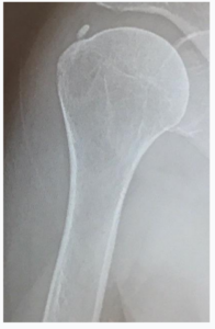
Imaging like an ultrasound, MRI, or radiograph (X-ray) plays a crucial role in the proper diagnosis and confirmation of calcific shoulder tendinopathy. X-ray images can offer a clear view of the calcific deposits impacting the shoulder or rotator cuff mobility, and may appear as a well-defined shadow or a light shadow, depending on the density of the calcification. Often physicians may complement an X-ray with an MRI as well as a diagnostic ultrasound examination. Ultrasound imaging allows a physician to perform a dynamic and thorough examination of the shoulder tendons.
How has calcific shoulder tendonitis traditionally been treated?
Fortunately, many treatment options exist to help treat the condition, alleviate pain, and improve function. Historically, patients experiencing calcific shoulder tendinopathy have been treated with a variety of methods, including:
- Nonsurgical treatments (rest, physical therapy, and anti-inflammatory medications)
- Ultrasound-guided needle aspiration and lavage
- Extracorporeal shock wave therapy
- Surgical treatments (i.e., tissue resection via open or arthroscopic surgery)
Non-surgical treatments can be helpful as a first line of treatment for most patients but could be insufficient when they offer only short-term relief. When these first-line treatments are unsuccessful, physicians may offer alternative treatments or refer patients to surgery. Alternative treatments may at times not be covered by insurance and surgical treatments may result in added downtime, cost, and morbidity.
A relatively new, minimally invasive procedure performed under ultrasound image guidance is now available to treat patients with calcific shoulder tendinopathy. The procedure is performed using the TenJet device and may offer a more reliable treatment compared to current non-surgical methods. It requires less downtime compared to open or arthroscopic shoulder surgery.
How does TenJet treat calcific shoulder tendonitis?
The TenJet device has been specifically designed to treat tendinosis and calcification in tendons without injuring surrounding healthy tissue. TenJet’s patented design uses a pressurized, high-velocity jet of saline as a cutting tool to precisely resect calcifications and diseased tendinopathic tissue.
The procedure is performed in an outpatient setting, most often under local anesthesia. Physicians make a small 5 mm incision to guide the TenJet needle into the area of calcium deposit. Ultrasound image guidance is used to identify and visualize the diseased tissue before and during the procedure, guide the TenJet needle, as well as perform the resection. Sutures are generally not needed to close the incision.
Is physical therapy required after the procedure?
The treating physician will guide their patient’s immediate post-treatment recovery and rehabilitation to help the patient heal and regain the strength in the shoulder. This plan may include physical therapy and home exercises.
- Depending on physician preference, patients may begin recovery in a sling for a maximum of 5 days post-procedure.
- Patients are advised to perform daily pendulum exercises and limit overhead lifting to less than 5 pounds for 14 days. However, patients may demonstrate improved mobility and may progress to light exercises within 5-14 days post-procedure.
- Within 4-6 weeks, most patients are expected to return to normal daily and light sports activity with minimal to no pain.
- Patients may continue to experience improvement (less pain, improved function) 3-4 months post procedure as they resume normal and advanced physical activity.
The recovery process for an ultrasound-guided tenotomy procedure using TenJet may be shorter and less painful than traditional open or arthroscopic tenotomy procedures. Individual patient results may vary depending on a variety of factors, but most patients experience significant relief following the procedure.
Calcific shoulder tendonitis can be painful. Treatment doesn’t have to be.
For any patient experiencing shoulder pain due to calcific tendinopathy of the shoulder, simply ignoring the problem is not a feasible solution. While many treatment options exist to alleviate pain and stiffness associated with calcific shoulder tendinopathy, an ultrasound-guided procedure using TenJet represents an excellent minimally invasive treatment option. Upon consideration of all diagnostic findings, physicians will be able to determine if a treatment using TenJet is the best option for their patient.
Please click on the following links for additional resources
TenJet Patient Information
TenJet Physician Finder
TenJet Physician Information
TenJet Shoulder Rehabilitation Webinar – Dr. Jason Genin
TenJet for Shoulder Tendinopathy Webinar – Dr. Jason Genin